


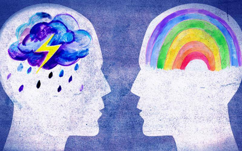
Creating colorful Pathways to happiness and swift recovery.
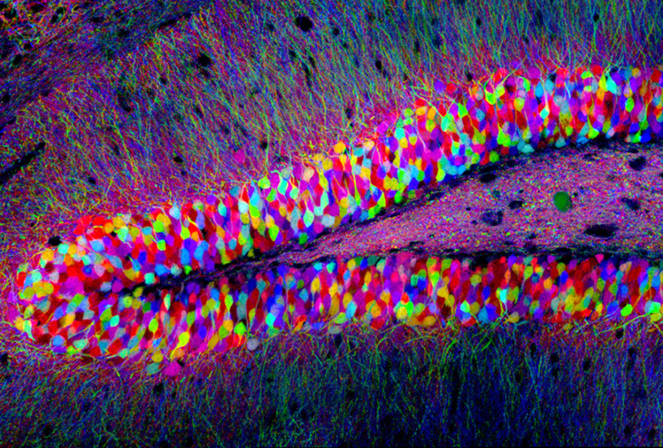
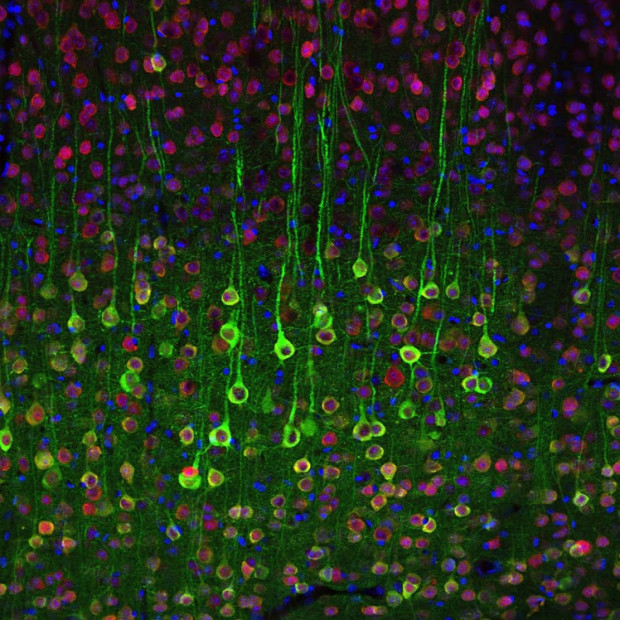
Brainbow is a process by which individual neurons in the brain can be distinguished from neighboring neurons using fluorescent proteins. By randomly expressing different ratios of red, green, and blue derivatives of green fluorescent protein in individual neurons, it is possible to flag each neuron with a distinctive color. This process has been a major contribution to the field of neural connectomics.
The technique was originally developed in 2007 by a team led by Jeff W. Lichtman and Joshua R. Sanes, both at Harvard University. With the recent advent of Brainbow in neuroscience, researchers are now able to construct specific maps of neural circuits and better investigate how these relate to various mental activities and their connected behaviors (i.e. Brainbow reveals information about the interconnections between neurons and their subsequent interactions that affect overall brain functionality). As a further extrapolation of this method, Brainbow can therefore also be used to study both neurological and psychological disorders by analyzing differences in neural maps.
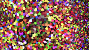
While earlier labeling techniques allowed for the mapping of only a few neurons, this new method allows more than 100 differently mapped neurons to be simultaneously and differentially illuminated in this manner. This leads to its characteristic multicolored appearance on imaging, earning its name and winning awards in science photography competitions
Art Meets Neuroscience
Below are pictures of what scientist see using the brainbow technique